
RQP74018 | |
| I. Background | |
| This PD-1 Cell Line is used to measure the binding of PD-1 to PD-L1/PD-L2. Programmed Cell Death Protein 1 (PD-1), a receptor expressed on activated T cells, binds to its ligands, PD-L1 and PD-L2, to negatively regulate immune responses. The PD-1 ligands are found on most cancers, and PD-1:PD-L1/2 interaction inhibits T cell activity and allows cancer cells to escape immune surveillance. The PD-1:PD-L1/2 pathway is also involved in regulating autoimmune responses, making these proteins promising therapeutic targets for a number of cancers, as well as multiple sclerosis, arthritis, lupus, and type I diabetes. | |
| II. Description | |
| Recombinant Jurkat T cell expressing firefly luciferase gene under the control of NFAT response elements with constitutive expression of human PD-1 (Programmed Cell Death 1, PDCD1, SLEB2, CD279, GenBank Accession #NM_005018). | |
| III. Introduction | |
| Host Cell: | Jurkat |
| Expressed gene: | NFAT-Luciferase-PD1 |
| Stability: | 32 passages |
| Synonym(s): | Programmed cell death 1, PDCD1, PD-1, PD1, SLEB2, CD279, HPD-L, PD1/NFAT, PD-1 NFAT, PD-1/NFAT |
| Freeze Medium: | 90% FBS+10% DMSO |
| Culture Medium: | 1640+10%FBS+300ug/ml hygromycin+1ug/ml puromycin |
| Mycoplasma Testing: | Negative |
| Storage: | Liquid nitrogen |
| Application(s): | Use this PD-1 Cell Line to screen for activators or inhibitors of PD-1 signaling in a cellular context • Characterize the biological activity of PD-1 and its interactions with ligands |
| IV. Description of Host Cell Line | |
| Organism: | Homo sapiens, human |
| Tissue: | Peripheral blood |
| Disease: | Acute T cell leukemia |
| Morphology: | Lymphoblast |
| Growth Properties: | Suspension |
| Ⅴ. Representative Data | |
 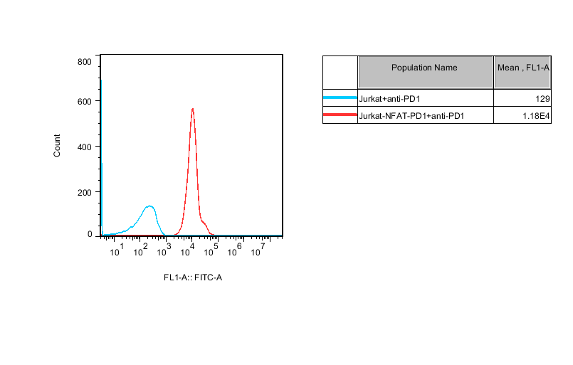 | |
Figure 1.Recombinant Jurkat T cell expressing firefly luciferase gene under the control of NFAT response elements with constitutive expression of human PD-1 (Programmed Cell Death 1, PDCD1, SLEB2, CD279, GenBank Accession #NM_005018). | |
| |
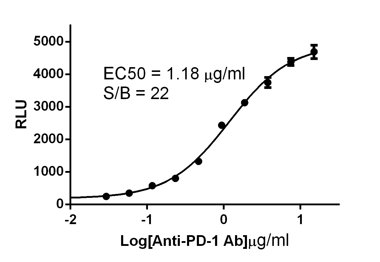
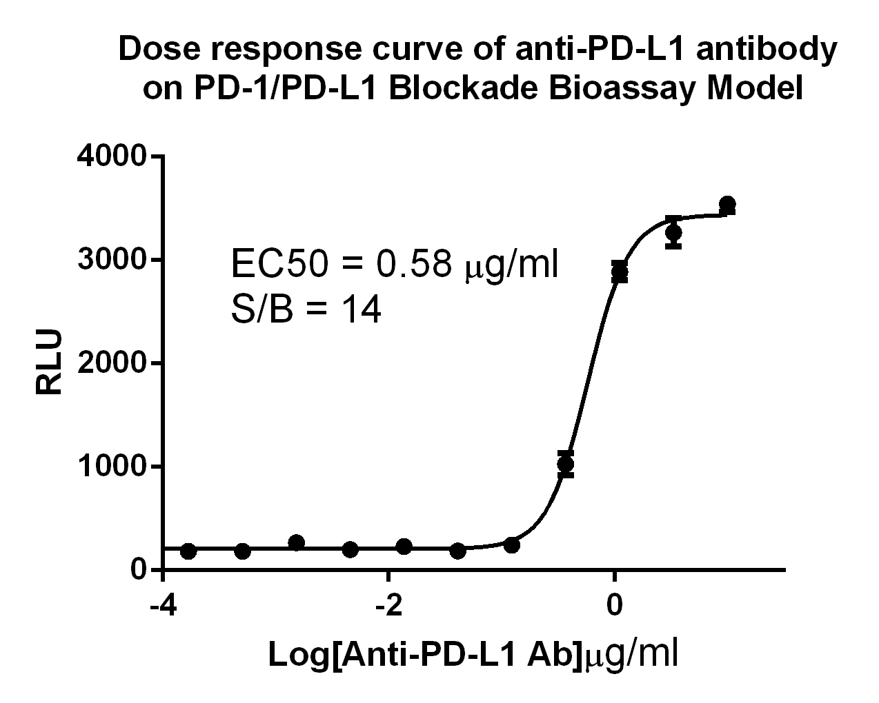
Figure 2. Dose response curve of anti-PD-1 antibody on PD-1/PD-L1 Blockade Bioassay Model. The bioassay consists of two genetically engineered cell lines, Jurkat_PD-1_NFAT-Luc Cells and CHO/TCR activitor/PD-L1 Cells. When co-cultured, the PD-1/PD-L1 interaction inhibits TCR-mediated luminescence. When the PD-1/PD-L1 interaction is disrupted, TCR activation induces luminescence (via activation of the NFAT pathway) that can be detected and quantified using Bio-Glo™ Reagent. | |
E-mail:sales@reqbio.com
Add:3rd Floor, No. 6, Lane 222,
Guangdan Road, Pudong New Area,
Shanghai,China

WeChat public account
